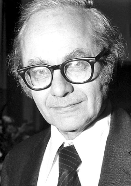
David Hunter Hubel
 Great Britain
Biography of David Hunter Hubel
David Hunter Hubel, an American neurophysiologist, was born in Windsor, Ontario, Canada to Elsa M. (Gunter) and Jess G. Hubel, who were of American origin. In 1929, the family moved to Montreal, where David developed an interest in science from his father and became fascinated with chemistry and electronics during his youth. He also showed musical abilities and devoted a significant amount of time to music from the age of five. From 1932 to 1944, he attended Strathcona Academy in Outremont. In 1944, Hubel enrolled in McGill University, where he graduated with honors in mathematics and physics. After obtaining his bachelor's degree in 1947, he entered the medical school at the same university, despite lacking knowledge in biology.
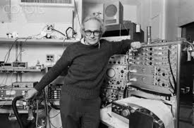
During summer breaks, Hubel worked at the Neurological Institute in Montreal, studying the nervous system. In 1951, after receiving his medical degree, he began studying clinical neurology first at the Neurological Institute for two years, and then at Johns Hopkins University in Baltimore, Maryland in 1954. The following year, Hubel was drafted into the U.S. Army and assigned to the Neurophysiology Department of the Walter Reed Army Institute of Research in Washington, D.C. It was there that he developed a tungsten microelectrode, a device used to record the electrical impulses of nerve cells. By implanting it in the brains of cats for specific periods of time, he could record the spontaneous activity levels of nerve cells in both sleeping and awake animals.
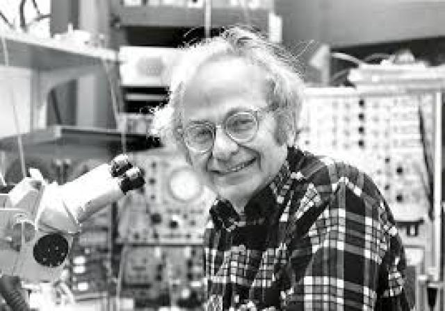
After his discharge from the army in 1958, Hubel continued his research at the Vernon Mountcastle laboratory at Johns Hopkins University. Mountcastle's laboratory was a recognized center for studying the sensory zones of the cerebral cortex. However, due to the outdated equipment in the lab, Hubel joined the research group of Stephen Kuffler, working at the Wilmer Institute and Johns Hopkins University. Kuffler, a leading expert in visual neurophysiology, studied the nerve activity (or microelectrical potentials) of retinal ganglion cells in cats. He discovered that retinal ganglion cells responded to light contrasts but not to uniform illumination. Kuffler also described the receptive fields of the retinal area, which showed changes in spontaneous nerve cell activity when stimulated. He found that retinal ganglion cells were either excited or inhibited when light was shone on their corresponding receptive field centers. Hubel and his colleague Torsten Wiesel aimed to study the receptive fields of other nerve cells in the visual analyzer, particularly those located in the visual centers of the cerebral cortex. The visual centers are one of the many functional areas of the cerebral cortex where higher cognitive processes like memory and perception take place. The visual analyzer begins with the photoreceptor cells (light-sensitive cells) in the retina, namely rods and cones. The nerve endings of the rods and cones project onto other cells in the retina, and the nerve impulses then travel through the optic nerves to the lateral geniculate body, from which they are transmitted to the visual centers of the cerebral cortex. The visual centers, consisting of millions of nerve cells arranged in layers, decode the nerve signals originating in the retina.
One of the first observations made by Wiesel and Hubel greatly expanded our understanding of how the nervous system functions. They inserted microelectrodes into the visual centers of the cat's cerebral cortex and recorded the spontaneous nerve cell activity. Attempting to elicit nerve cell activity in the cortex, they tested various visual stimuli. Accidentally moving the microscope stage glass slide across the receptive field of the nerve cell into which the microelectrode was implanted, Hubel noticed that the cell started discharging. Initially, the researchers were puzzled by this phenomenon, but they soon realized that the cell was responding to a strip of light on the glass slide. While Kuffler's retinal cells responded to circular "patterns," the nerve cells in the visual area of the cerebral cortex responded to linear light stimuli. In 1959, Kuffler became a professor of pharmacology at Harvard Medical School in Boston, Massachusetts, and his research group, including Hubel and Wiesel, moved there. In 1964, the Department of Neurobiology was established at Harvard under the leadership of Kuffler. In 1967, Hubel became the department chair, and the following year, he was appointed as a professor of physiology. During their time at Harvard, Hubel and Wiesel conducted numerous experiments in which they inserted microelectrodes into the visual centers of live cats and monkeys, recording the spontaneous activity of the nerve cell into which the microelectrode was implanted. They hoped to stimulate the receptive fields of the retina with linear light perception under various orientation angles until they found the most effective stimuli for a group of cells along the electrode path. Sometimes, they inserted the electrode perpendicular to the brain surface, and in other experiments, they inserted it at an angle. After sacrificing the experimental animals, they compared the data on nerve tissue activity and its anatomical features. The researchers also developed a method for introducing radioactive substances into the eyeball. By following the fibers of the optic nerve from the retina to the visual centers, these labeled substances helped provide an understanding of the anatomical features of the visual centers of the brain.
As a result of their experiments, Hubel and Wiesel discovered that the visual centers of the cerebral cortex are organized in the form of periodic vertical complexes, which they called dominant ocular columns and orientation columns. In these columns, nerve cells process the necessary reframing of information transmitted from the retina to the visual centers. Dominant columns combine neuronal impulses from both eyes, while orientation columns transform circular receptive fields of the retina and lateral geniculate bodies into linear receptive fields. Hubel and Wiesel found that this information processing involved a hierarchy of simple, complex, and hyper-complex nerve cells, which functioned according to the principle of increasing or progressive convergence. The principle of progressive convergence explains how complete images can be created from numerous individual bits of information received from retinal neurons in the visual area of the cerebral cortex. Other functional centers of the cerebral cortex may be organized in a similar manner. Hubel and Wiesel's research in visual neurophysiology had a significant impact on clinical ophthalmology, particularly in the treatment of congenital cataracts. For instance, they discovered that such cataracts should be removed in early childhood to preserve vision.
Hubel and Wiesel were awarded half of the 1981 Nobel Prize in Physiology or Medicine "for their discoveries concerning information processing in the visual system." The other half of the prize was awarded to Roger W. Sperry. In his presentation speech, David Ottoson from the Karolinska Institute reminded the audience that Hubel and Wiesel demonstrated "how different parts of the image on the retina are read and interpreted by cells in the cerebral cortex... The cells are arranged in columns; the analysis takes place in a strict sequence from one cell to another, and each nerve cell is responsible for a particular detail in the overall picture." Ottoson emphasized that the researchers also found that the ability to decode information from the retina develops soon after birth. This finding is crucial because the inability to see with even one eye "even for a few days during this period... can lead to permanent visual impairment." Hubel married Shirley R. Izard in 1953, and they had three sons.
Hubel received an honorary doctorate from McGill University and was awarded the Lewis Rosenstiel Medal for Distinguished Work in Basic Medical Research at Brandeis University (1972), the John F. Friedenwald Memorial Award from the Association for Research in Vision and Ophthalmology (1975), the Karl Spencer Lashley Award from the American Philosophical Society (1977), the Louise GrossHorwitz Prize from Columbia University (1978), the Dixon Prize in Medicine from the University of Pittsburgh (1979), and the George Ledlie Prize from Harvard University (1980). He was a member of the National Academy of Sciences and the American Academy of Arts and Sciences.


